

How To Determine Resolution Regeneration Repair
Introduction
When exposed to trauma, infection, thermal or chemic injury, or other agin stimuli, all organisms including humans evoke an immediate, programmed, not-antigen specific allowed response to preserve the organism's integrity and re-establish homeostasis (Medzhitov, 2008). This reaction is governed by cells of the innate immune organization and defines the acute inflammatory response (Mosser and Edwards, 2008; Chen and Nuñez, 2010). Acute inflammation is the offset stage of healing of all tissues, and normally results in repair and regeneration of the damaged structures (Mantovani et al., 2022). If the sequence of events comprising tissue healing is interrupted or dysregulated, the typical healing of host tissue becomes impaired (Gerstenfeld et al., 2003). Furthermore, if the injurious stimulus is not quickly mitigated, either the organism equally a whole will succumb (if the injury or resulting response is overwhelming), or the local tissues may progress to a state of chronic inflammation, in which ongoing injury and attempts at repair persist. Thus, the stop result of an agin stimulus may vary from complete restoration of anatomical form and function at one end of the spectrum, to subsequent expiry at the other extreme; injuries often upshot in intermediate outcomes including partial tissue regeneration, fibrosis, and/or chronic inflammation.
With regards to bone and soft tissues, the response to injury is no different than for other organs. When bone is subjected to trauma or an adverse stimulus, the resident cells release numerous cytokines, chemokines, and other substances that initiate local vasodilatation and efflux of inflammatory cells from the circulation; a pro-inflammatory pour of events is launched to terminate the adverse upshot and initiate the regenerative process (Marsell and Einhorn, 2022; Karnes et al., 2022). Although, numerous cells are directly involved in these ongoing activities, local macrophages, as well as circulating surveillance monocyte/macrophages orchestrate the ensuing serial of biological events (Fujiwara and Kobayashi, 2005; Medzhitov, 2008; Nich et al., 2022; Sinder et al., 2022; Kaur et al., 2022). Specialized cellular systems have evolved including design recognition receptors (PRRs) to identify chemical motifs from bacteria and infectious agents (so chosen pathogen-associated molecular patterns or PAMPs) and byproducts of cell death and tissue injury (damage-associated molecular patterns or DAMPS) (Kawai and Akira, 2010). When PRRs are ligated, a system of effector mechanisms including Toll Like Receptors (TLRs), Nucleotide-bounden Oligomerization Domain (NOD) and leucine-rich repeat-containing receptors (NLRs), Retinoic acid inducible gene (RIG) receptors, and others transmit these signals through intermediate molecules to upregulate the germination and release of pro-inflammatory substances (Akira and Takeda, 2004; Medzhitov, 2008). These include cytokines e.g., tumor necrosis factor alpha (TNFα), Interleukin i beta (IL-1β), IL-vi, IL-8, and others, chemokines including macrophage chemotactic protein 1 (MCP-1), macrophage inhibitory poly peptide 1 (MIP-one), reactive oxygen intermediates (such as inducible nitric oxide synthetase or iNOS), and growth factors (such every bit vascular endothelial growth cistron or VEGF, transforming growth factor beta or TGFβ etc.). These cells and substances eradicate invading microbes, limit the injurious stimulus, and recruit more cells to participate in the biological confrontation, and begin the resolution and reparative phases (Medzhitov, 2008; Mosser and Edwards, 2008; Mantovani et al., 2022).
This paper will summarize the important biological processes of inflammation as they chronicle to bone healing and emphasize the disquisitional intercellular communications that participate in repair of os subjected to agin stimuli. In vitro and in vivo inquiry performed in our laboratory and by others that facilitates bone repair in inflammatory conditions will be highlighted.
The Biological Reaction to Wear Particles From Joint Replacements: A Image for Acute and Chronic Inflammation
Traditionally, nigh joint replacements have used a begetting couple composed of ultra-high molecular weight polyethylene (UHMWPE), and a metal or ceramic counter surface. This bearing couple has recently been improved, with the development of enhanced crosslinking of the polyethylene and embedded anti-oxidants, and past reducing the surface asperities and polishing of the countersurface. However, for the get-go twoscore years of articulation replacement surgery, the biological reaction to wear particles, and the resultant sterile inflammation and os loss (known as periprosthetic osteolysis) were the predominant reasons for revision (redo) surgery (Jacobs et al., 2001; Purdue et al., 2007; Gallo et al., 2022) (Figure one). This discipline has been studied extensively by our grouping and others; numerous in vitro and in vivo models have been developed to simulate the events of article of clothing particle-induced inflammation.
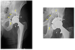
Effigy i. Periprosthetic osteolysis postal service total hip replacement (THR). The left radiograph shows a hybrid THR with a cemented stalk and a cementless cup with screws. The components are well stock-still, however, in that location is polyethylene wearable and the metallic loving cup has fractured next to the screw holes (annotation: ii of the spiral holes are larger than they should be and confluent instead of separate—black arrow). The small white radio-dense particles correspond metallic debris from the cup (white arrows). At that place is a large radiolucent blackness area of bone destruction (osteolysis) (yellow arrows) surrounding the acetabular component. The radiograph on the right is a magnified view of the acetabular area.
In general, wear particles stimulate a non-specific macrophage dominated inflammatory reaction characteristic of the innate allowed system, in a background fibrovascular stroma (Goodman et al., 1998; Goodman, 2007). The characteristics of the wear particles are important to this reaction: smaller (0.three to <5–10 μm) irregularly shaped particles of polymers announced to be more inciting of an inflammatory response, compared to ceramic or metallic particles, all the same this point is controversial (Goodman, 1994; Kaufman et al., 2008; Goodman et al., 2009). Particles ~i μm or less are the nearly prominent and reactive ones (Campbell et al., 1995). In addition, to the above particle characteristics, the surface expanse, surface energy and overall number and volume of particles are cardinal factors in the resultant histological reaction (Shanbhag et al., 1994; González et al., 1996; Green et al., 1998, 2000). Certain metal particles and byproducts can stimulate both the innate and adaptive immune systems, the latter occurring when the metallic moiety and attached protein office as a hapten (Haynes et al., 1993; Hallab et al., 2001; Caicedo et al., 2008). Indeed, all particles are spring to serum proteins such equally albumin, alpha-one-antitrypsin, apolipoprotein, and others, and activate specific cell surface receptors to engage the inflammatory cascade (Nakashima et al., 1999; Sun et al., 2003). These complexes are recognized past prison cell surface receptors, or if small enough, phagocytosed altogether (Nakashima et al., 1999; Purdue et al., 2007). Although, numerous biological pathways in macrophages, fibroblasts and other cells are involved in these events, the cardinal molecules involved in particle-associated inflammation include the adapter protein Myeloid Differentiation chief response gene 88 (MyD88), and the transcription factor nuclear factor kappa-calorie-free-chain-enhancer of activated B cells (NFκB) (Nakashima et al., 1999; Clohisy et al., 2004; Ren et al., 2004; Baumann et al., 2005; Pearl et al., 2022). Activation of MyD88 and NFκB atomic number 82 to the transcription of numerous pro-inflammatory substances and upregulation of the innate and (to a lesser degree with respect to wearable particle illness) the adaptive allowed systems (Pearl et al., 2022; Landgraeber et al., 2022; Nich et al., 2022). In bone and the surrounding tissues, this results in an influx of primarily monocyte/macrophages, only as well mast cells, polymorphonuclear leukocytes, T lymphocytes, osteoclasts and other cells are present (Hallab and Jacobs, 2022). The resulting pro-inflammatory environment leads to increased bone destruction by cells of the monocyte/macrophage/osteoclast lineage and suppressed bone formation past cells of the mesenchymal stem jail cell (MSC)/osteoblast lineage (Kadoya et al., 1996; Vermes et al., 2000; Jacobs et al., 2001). With regards to osteoclastogenesis, the Receptor Activator of Nuclear Factor-kappa B Ligand (RANKL)-RANK- osteoprotegerin (OPG) axis becomes dysregulated, leading to increased osteoclast formation and activation (Haynes et al., 2001). Furthermore, soluble and particulate cobalt-chrome molybdenum alloy (and other particle types) are capable of activating the intracellular inflammasome pathway which increases the secretion of IL-one and other pro-inflammatory cytokines (Caicedo et al., 2008). As more wear particles are continuously produced with use of the artificial implant, the acute inflammatory reaction becomes chronic, with progressive synovitis and bone destruction. In addition, the presence of endotoxin on the particles and other bacterial byproducts tin can sustain and exacerbate the inflammatory reaction (Bi et al., 2001).
In vitro and in vivo Models of Particle-Induced Inflammation Advise Potential Avenues for Handling
In general, our tact has been to develop in vitro models for proof-of-principle testing of new concepts and biologics, then expand and validate these hypotheses using in vivo models that simulate the biological events of wear particle disease as closely equally possible. One appreciates the associated temporal compression of such models compared to a disease in humans that commonly takes many years to develop. Moreover, in investigating the resultant inflammatory os loss associated with wear particles, one besides recognizes the suppressive effects of particles on MSC-osteoblast lineage cells (Wang et al., 2002; Chiu et al., 2006, 2009; Goodman et al., 2006; Ramachandran et al., 2006; Atkins et al., 2009; Pajarinen et al., 2022a). This realization has led to novel methods not but to mitigate bone destruction, but to raise bone formation, subjects very relevant to the broader topics of tissue engineering and repair of bone. It is likewise recognized that the pro-inflammatory effects associated with wear particles are not the merely factors leading to dysregulated bone biological science around joint replacements; other factors include the presence of bacterial ligands, mechanical forces, fluid pressure, and immune reactions especially to metallic byproducts etc. (Aspenberg and Herbertsson, 1996; Aspenberg and Van der Vis, 1998; Bi et al., 2002; Cho et al., 2002; Choi et al., 2005; Caicedo et al., 2008; Greenfield and Bechtold, 2008).
Numerous studies have established that wear particles both upregulate the inflammatory cascade and suppress the pathways that facilitate os germination (Jacobs et al., 2001; Goodman, 2007; Purdue et al., 2007; Goodman and Ma, 2010). In vivo models of particle induced osteolysis take the difficulty of simulating a complex series of biological events in a short catamenia of time, in a cost-effective and practical manner. Yet, both small and large brute models have demonstrated some of the important pathogenetic mechanisms leading to particle-associated osteolysis (Lind et al., 1998; Cordova et al., 2022; Moran et al., 2022).
Originally, our laboratory used simpler models encompassing a single bolus of unlike particles alone, or with more basic implants resurfacing but one side of a joint, or in bone harvest chambers in rabbits; we also implanted particles around a solid intramedullary rod in mice (Goodman et al., 1993; Goodman, 1994; Sacomen et al., 1998; Epstein et al., 2005b; Zilber et al., 2008). While these models provided important information regarding the astute inflammatory reaction to particles (which perhaps was more than relevant to the bedding in phase of wear and osseointegration of implants), there were several deficiencies. Offset, particles are continuously produced from bearing surfaces in man joint replacements and a single bolus of particles does not simulate this scenario. Second, the cellular processes reflective of more chronic particle exposure and the longer-term attempts at re-establishment of tissue homeostasis could not be investigated. Tertiary, some of the models, such as the calvarial model (using a flat os) were anatomically and physiologically dissimilar from the clinical situation in which human implants are placed in long bones that have a unlike anatomical and biomechanical construction, and blood supply. Furthermore, the calvarial model does not ordinarily use an implant to simulate a prosthesis. Fourth, the rabbit models were expensive and proved difficult to use with cut-border technologies such equally genetic manipulation of cells, advanced imaging techniques etc. Nevertheless, single bolus models are still relevant, equally they have demonstrated that wear particles of unlike materials stimulated a macrophage dominated foreign body inflammatory reaction that increased os destruction and diminished bone formation. The key pro-inflammatory cytokines (TNFα, IL-1β, IL-vi, and others) and chemokines (MCP-one, etc.) associated with this reaction were identified (Trindade et al., 1999; Epstein et al., 2005a,b). Using these models, we investigated potential treatments for osteolysis, such as the furnishings of oral non-steroidal anti-inflammatory medications, an oral p38 mitogen-activated protein kinase (MAPK) inhibitor, and locally placed growth cistron (due east.g., Transforming Growth Gene beta) (Goodman et al., 1999; Kumagai et al., 2008). Still, these substances also adversely affected bone formation, and the timing of delivery and optimal dosage were hard to found in vivo.
As a consequence, more than representative models of continuous particle delivery over a more extended time period in small rodents were developed by our laboratory. These models were less costly, fake the clinical scenario more closely, and could have advantage of newer genetic and imaging technologies. Thus, we adult the murine femoral continuous intramedullary particle infusion model, in which a improvidence pump implanted in the subcutaneous paraspinal region was connected via tubing to a hollow titanium rod placed in the intramedullary canal of the distal third of the femur (Figure 2). Particles and potential therapeutic agents could exist loaded into the pump and continuously delivered into os via the hollow rod over ~28 days. The model was validated start ex vivo, prior to its apply in live animals (Ortiz et al., 2008a,b). Using histomorphometry, immunohistochemistry, and microCT analysis, we and then reported that continuous infusion of clinically relevant polyethylene particles produced a chronic inflammatory macrophage dominated reaction and decreased local bone book, compared to infusion of the carrier alone (Patterson et al., 2008). Recently we demonstrated that extending the particle delivery time up to 56 days leads to further evolution of chronic inflammation, with continued macrophage activation and bone loss, similar to the progressive clinical scenario (Pajarinen et al., 2022b). We also extended this model to study systemic macrophage trafficking by injecting genetically altered reporter macrophages into the tail vein immediately subsequently surgery, and repeatedly tracked the migration of these cells throughout the body not-destructively via bioluminescence (Ren et al., 2010). To follow systemic trafficking of reporter MSCs, nosotros needed to develop another technique, using left ventricular cardiac jail cell injection in the beating heart, because the much larger MSCs delivered through the tail vein would sequester in the pulmonary microvasculature, rather than pass through the lungs into the arterial organization (Fritton et al., 2022). From these experiments nosotros learned the following: (a) infusion of the chemokine MCP-1 or polyethylene particles via the osmotic pump induces systemic recruitment of reporter macrophages to the local area which results in osteolysis. This macrophage reporter cell trafficking and bone loss could be mitigated by interrupting the MCP-one-CCR2 chemokine-receptor axis using an MCP-ane receptor antagonist or reporter cells from knockout mice that do not possess the CCR2 receptor (CCR2− cells) (Gibon et al., 2022a); (b) luciferase expressing reporter MC3T3 pre-osteoblasts injected into the left ventricle migrated systemically to the area of particle infusion in the distal femur and were associated with increased bone mineral density and markers of bone turnover locally. These effects could exist mitigated by injection of an inhibitor of the C-C chemokine receptor CCR1, which interferes with both leukocyte and MSC chemotaxis (Fritton et al., 2022; Gibon et al., 2022b). The above interventions revealed the local and systemic pathways associated with particle-associated inflammation and suggested potential mechanistic interventions for treatment.
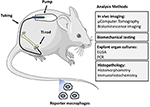
Effigy ii. The murine femoral continuous intramedullary particle infusion model. First, the osmotic pump is loaded with biomaterial wear droppings and and then implanted in the subcutaneous tissue at the dorsum of the mouse. The pump is then connected via subcutaneous tubing to a hollow titanium rod that has been printing fit into the intramedullary canal of the distal femur. This arrangement facilitates continuous delivery of biomaterial wear debris to the intramedullary space for 28 days, resulting in continued low class inflammation and os loss. The particle delivery can be further extended by changing the pump in a minor surgery. The resulting bone loss at the distal femur can exist quantified past imaging techniques such as μCT, biomechanical testing of the peri-implant bone, and histomorphometry. The chronic inflammatory reaction tin can be quantified by assay of femoral explant cultures and various histopathological techniques including identification of specific jail cell populations and their activation states past immunohistochemistry. Finally, systemic homing of macrophages and other cells to the surface area of inflammation tin can be quantified by utilizing luciferase labeled reporter macrophages that are injected into the circulation via the tail vein. Adding biologics to the pump with the particles allows the study of potential therapeutic effects of dissimilar locally infused treatments.
More than recently, we have engaged 3 strategies to decrease particle associated bone devastation using our murine models (Figure iii). First, we have coated the distal femoral intramedullary rod with a mutant MCP-i (MCP-i is also referred to every bit CCL2) poly peptide chosen 7ND recombinant protein via a layer-by-layer (LBL) technique to office every bit a drug eluting device to decrease macrophage trafficking locally (Keeney et al., 2022). Using microCT, immunohistochemical staining, and bioluminescence imaging, local delivery of 7ND protein via the LBL blanket decreased systemic reporter macrophage recruitment to the particle infusion area, decreased the number of osteoclasts locally, and mitigated wear particle-induced bone loss in the distal femur (Nabeshima et al., 2022).
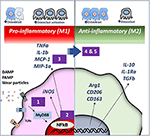
Effigy iii. Strategies for immunomodulation to mitigate periprosthetic osteolysis induced by wear particles. Wear particles and adherent pathogen-associated molecular patterns (PAMPs) and byproducts of jail cell death and tissue injury (impairment-associated molecular patterns or DAMPS) can exist recognized by Price-like Receptors (TLRs) and other receptors on macrophages, which then actuate downstream pathways including the key transcription cistron Nuclear Factor-kappa B (NFκB). The induced pro-inflammatory responses driven by NFκB activation include the expression of inducible nitric oxide synthetase (iNOS) and cytokines/chemokines including Tumor Necrosis Gene alpha (TNFα), Interleukin one beta (IL-1b), Macrophage Chemotactic Protein 1 (MCP-1), Macrophage Inhibitory Protein i alpha (MIP-1α), and others. These events may atomic number 82 to periprosthetic osteolysis due to reduced osteoblast and increased osteoclast action. We have demonstrated that the particle-induced osteolysis can be mitigated by inhibiting (i) the TLR pathway; (2) NFκB activation; or (3) macrophage migration using a mutant MCP-1 called 7ND recombinant poly peptide. Alternatively, pro-inflammatory macrophages (M1, Left) can be polarized by (4) Interleukin 4 (IL-4) handling or (5) genetically modified or preconditioned mesenchymal stem cells (MSCs) (run across Figure 4 for details) into an anti-inflammatory, pro-tissue repair macrophage (M2) phenotype (Right). M2 macrophages are identified by their expression of Arginase 1 (Arg1) and the surface markers CD206 and CD163. M2 macrophages produce Interleukin 10 (IL-10), IL-1 receptor antagonist (IL-1ra), and Transforming Growth Factor beta (TGFβ). Myeloid Differentiation primary response 88 (MyD88) is a universal adapter protein that is downstream of near all TLRs (except TLR3), and leads to activation of NFκB.
Our second strategy was to interfere with the master transcription gene NFκB, which regulates the expression of pro-inflammatory cytokines and chemokines of the innate immune organisation, and if persistently activated, leads to decreased bone formation and increased bone destruction. Nosotros take achieved this downregulation of NFκB via local infusion of an NFκB decoy oligodeoxynucleotide (ODN), a synthesized duplex Dna that suppresses NFκB activity through competitive binding. Nosotros take confirmed the effectiveness of this strategy in in vitro studies, and in vivo, using the murine calvarial model and the femoral intramedullary particle infusion model (Lin et al., 2022, 2022a; Sato et al., 2022).
Our third strategy is to polarize local macrophages temporally, from an initial pro-inflammatory phenotype (also called M1) to an anti-inflammatory pro-regenerative (M2) phenotype. We accomplished this by exposing the M1 macrophages to interleukin-four, an anti-inflammatory cytokine. In vitro studies were get-go performed in co-civilisation of undifferentiated macrophages (M0), M1, or M2 together with pre-osteoblasts to determine the optimum time and concentration of cells and IL-4 to optimize bone formation. Polarizing M0 or M1 macrophages to M2 macrophages past the addition of IL-4 optimized matrix mineralization at 3 weeks, and osteocalcin and element of group i phosphatase expression, if the IL-4 was added after ~72 h (Loi et al., 2022b; Córdova et al., 2022). Adding IL-iv earlier or continuously was less optimal. This finding substantiated the belief that a given menses of inflammation and osteoprogenitor priming was necessary for optimizing os formation (Gerstenfeld et al., 2003). After further in vitro validation, we subsequently showed that local commitment of IL-4 poly peptide decreased the inflammatory response to particles, and increased cyberspace os formation using the calvarial and the femoral intramedullary particle infusion models (Nich et al., 2022; Pajarinen et al., 2022, 2022b; Sato et al., 2022).
Thus, 3 local potentially translational strategies for modulation of the innate allowed system in response to particle claiming were shown to mitigate the agin inflammatory response and augment bone germination. Although, wear particle disease involves both local, and to some degree, systemic activation of innate allowed processes, our group has focused on developing treatment options that are applied locally, direct to the site of the particle induced inflammation; this approach concentrates on altering the biological sequelae of particle disease directly at the source of the problem thereby limiting potential systemic toxicity of the treatments. These potential treatments might have a role in the early on stages of osteolysis, when the prosthesis is still salvageable. This biologically based approach supplements ongoing innovations in material scientific discipline and tribology of joint replacements.
Modulation of Inflammation: Relevance to Tissue Engineering and Os Healing
As stated previously, inflammation is the get-go stage of healing for all tissues. Interestingly, aging is associated with a land of ongoing low grade inflammation ("inflammaging"), and dysregulated macrophage polarization in response to potentially injurious stimuli (Mahbub et al., 2022; Gibon et al., 2022). In other words, with crumbling, an injury does not always effect in a measured coordinated inflammatory reaction with subsequent resolution and repair, but may develop into a chronic inflammatory state with ongoing tissue destruction. Furthermore, aging is associated with a general subtract in the response of both the adaptive and innate immune systems to adverse stimuli (Frasca and Blomberg, 2022). These facts may explain the delayed and/or insufficient healing in the elderly when subjected to traumatic injuries or other adverse stimuli including infectious disease.
The immune organisation and the musculoskeletal systems are intimately co-dependent (Loi et al., 2022a). Crosstalk between macrophages and other hematopoietic cells, and MSC lineage cells is important to hematopoiesis, immunomodulation, and the resolution of inflammation, as well every bit the healing and repair of musculoskeletal tissues (Maggini et al., 2010; Mountziaris et al., 2022; Guihard et al., 2022; Mantovani et al., 2022; Wu et al., 2022; Six et al., 2022; Loi et al., 2022b). We and others have shown that continuous crosstalk between macrophages and MSC lineage cells are critical to os healing (Mountziaris et al., 2022; Omar et al., 2022; 6 et al., 2022; Loi et al., 2022b). In addition, with crumbling, osteogenesis by MSC lineage cells is depressed; these effects have been shown by our group to exist associated with ongoing upregulated NFκB activity by anile MSCs (Lin T. H. et al., 2022). Thus, i potential approach to facilitating bone healing in the elderly might be local/regional modulation of NFκB activity in macrophages, directly or indirectly. This approach has been alluded to above.
Two additional approaches to immunomodulation by altering MSCs to improve bone healing in inflammatory clinical scenarios have been explored by our grouping (Figure 4). These approaches are potentially relevant to os repair in the young and aged alike.
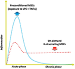
Figure iv. Modulating inflammation with specialized MSCs to raise bone formation. Optimal bone regeneration is mediated past a transient acute inflammatory reaction (for several days), followed by the resolution of inflammation and the tissue repair phase (bluish solid line). Harm or dysregulation of the acute inflammatory phase may lead to unresolved chronic inflammation and subsequent delayed bone healing (ruby-red dashed line). The strategy of using MSCs preconditioned by exposure to both lipopolysaccharide (LPS) plus TNFα ex vivo mimics the acute phase response and enhances the MSCs' osteogenic and immunomodulating abilities. Alternatively, genetically modified MSCs that first sense NFkB activation then over-limited IL-4 secretion ("on demand") can respond to unresolved chronic inflammation by modulating the local conditions into the desired anti-inflammatory, tissue repair environment for improved bone healing.
The beginning approach includes preconditioning the MSCs prior to their utilise, mimicking the inflammatory environs to which the MSCs are exposed when they showtime enter the area of tissue damage and regeneration. Other laboratories have demonstrated that preconditioning of MSCs by exposing them to inflammatory cytokines including interferon gamma (INFγ) and TNFα synergistically enhances their immunomodulatory properties by suppressing the activation of T cells (Ren et al., 2008; François et al., 2022). We developed a novel method of preconditioning of MSCs for bone healing applications, using a combination of lipopolysaccharide (LPS- a constituent found in the jail cell wall of gram negative bacteria) together with TNFα (Lin et al., 2022b). When these preconditioned MSCs (pMSCS) were co-cultured with macrophages, the macrophages polarized from an M1 to an M2 phenotype and were associated with increased osteogenic differentiation of the MSCs, and greater alkaline phosphatase expression and matrix mineralization. Given the fact that inflammation is oftentimes role of recalcitrant os infections, non-matrimony of fractures, periprosthetic osteolysis, osteonecrosis, and other diseases of bone, preconditioning of MSCs may have a directly translational awarding in the healing of astute and chronic bone defects. Furthermore, the preconditioning protocol adult in our laboratory may prove useful for immunomodulation of other systemic inflammatory disorders such as sepsis, rejection of solid organ transplants etc. in which MSCs are infused.
The 2nd approach encompasses genetic modification of MSCs to over-limited the immune-modulating pro-regenerative cytokine IL-4. We accept developed 2 constructs to accomplish this goal. In one construct, overexpression of IL-iv by MSCs is continuous; in the other construct, IL-4 is simply overexpressed by MSCs when NFκB activity is first sensed as upregulated (Lin et al., 2022c). In the latter construct, when NFκB activity diminishes, the excess production of IL-4 is stopped. Thus, when an inflammatory stimulus is encountered, these genetically modified MSCs (GM-MSCs) tin can secrete increased amounts of IL-4, subsequently polarizing M1 macrophages (in the vicinity) to an M2 phenotype. Because acute traumatic conditions or adverse stimuli require an initial pro-inflammatory surround to precondition or license the local MSCs for bone healing or other immunomodulatory functions, the IL-4 secreting MSCs would be about useful several days afterwards astute injury, or in chronic inflammatory conditions. The advantage of the NFκB sensing IL-4 overexpressing MSCs is that the commitment of IL-four could be temporally and spatially tailored to an ever changing inflammatory and immune environment, i.eastward., be context dependent.
Give-and-take
Acute and chronic inflammation are biological processes inside the immune system that are integral to the sustenance of life for all organisms. In humans, the innate and adaptive immune systems are highly adult. The sometime (innate immunity) responds to injury or adverse stimuli in a pre-adamant, non-specific way that is generally dependent on the interaction of cells with chemical motifs that incorporate the adverse stimulus. The latter (adaptive amnesty) is dependent on the interaction of specific receptors on cells (antigen presenting cells as well as T and B lymphocytes) with a more specific antigenic stimulus. Previously it was idea that only the adaptive allowed system had the potential for memory of a previously encountered stimulus challenge; it is now recognized that the innate allowed arrangement has a machinery that "remembers" previous interactions (Italiani and Boraschi, 2022). With subsequent challenges by the same or like stimuli, monocytes/macrophages can increment ("trained immunity") or decrease ("tolerance") the production of cytokines, chemokines, and other substances to effectively deal with a potentially injurious event (Dobrovolskaia and Vogel, 2002). This non-specific innate immune memory can last for months and allows monocytes/macrophages to attune their functional state according to the persistence of the adverse stimulus. This innate allowed memory optimizes survival of the organism by facilitating a relatively speedy and enhanced reaction to potentially harmful stimuli, but also allows a measured defensive response that does not consume the organism (Medzhitov et al., 2022). We are currently exploring these concepts, but much work remains in this image-changing research. For example, it may be possible to create implants that release specific substances based on local contextual cues (e.g., the presence of bacterial ligands or excessive amounts of habiliment debris); these released substances would then precondition local MSCs or other cells to undertake specific immunomodulatory activities.
How are the higher up concepts related to wear particle illness? Wear particles are continuously produced by orthopedic implants with repeated usage. In full general, debris from normally used polymers, ceramics and metals in orthopedics provoke an innate immune response; in some cases, protein-metallic byproducts can also human activity as haptens, thereby stimulating the adaptive immune system as well. Thus, the biological reaction to article of clothing particles from orthopedic implants can office equally a paradigm for exploring the mechanisms associated both with acute and chronic inflammation and activation of the immune system using relevant in vitro and in vivo models. Once these biological processes are elucidated, information technology may be possible to (a) optimize the composition and pattern of biomaterials and implants, and (b) attune tissue-implant responses to facilitate integration of the device or otherwise improve its function in vivo in the short and long terms. For articulation replacements specifically, these concepts tin can be translated from bench to beside. For example, implants could exist coated with biological substances to facilitate and even expedite initial osseointegration and promote early physiological loading, thus providing pathways for earlier return to function. Methods to mitigate infection, one of the leading causes of implant failure, need to be addressed. This might exist accomplished using newer fabrication (for example 3D press) and coating techniques; alternatively, the periprosthetic environment could be manipulated immunologically to minimize bacterial colonization and expansion. The techniques above to enhance osseointegration and prevent infection could exist combined with novel methods to interrogate and sense the periprosthetic environment and so release specific diagnostic and therapeutic agents on need. These and other interventions would require all-encompassing in vitro and in vivo testing using relevant fauna models. Many of the immune modulating interventions discussed higher up have merely been delivered in the brusque term. Longer term studies outlining strategies for resolving inflammation at an appropriate time indicate, without local or systemic agin effects are needed. Furthermore, novel strategies are needed to address continuous particle production and chronic inflammation over many decades, and potential methods to facilitate particle clearance. Indeed, continuous long-term immunomodulation may have deleterious furnishings to the host. Thus, solutions will undoubtedly entail better methods of diagnosis of particle-associated inflammation including potential biomarkers that are more than sensitive than conventional radiographs, computed tomography, or MRI. In this way, biological interventions could be delivered intermittently at timepoints of college particle loads and inflammatory responses.
An understanding of the constant interactions amidst cells of the monocyte-macrophage-osteoclast lineage and the MSC-osteoblast lineage also is critical to tissue applied science of os. Indeed, the processes of inflammation and bone and soft tissue healing are and then intertwined, that damage of ane procedure impacts the other (Guihard et al., 2022; Mantovani et al., 2022; Loi et al., 2022a,b). Thus, in that location are significant opportunities for modulating inflammation to obtain a desired outcome for bone healing and regeneration (Mountziaris et al., 2022).
By studying vesture particle affliction and related pathologies of bone, our group and others have begun to understand the cellular and molecular processes associated with inflammation and activation of the innate immune system and in item, their office in the formation and destruction of bone. This understanding has led to the design of innovative in vitro and in vivo models to simulate the activities of the innate immune organisation and develop potential local treatments to mitigate injurious stimuli and facilitate bone maintenance and repair. Every bit the crosstalk between the innate immune system and MSCs is so critical to os and soft tissue modeling, investigating ways to optimize their communications has been a continued focus of our current investigations.
On a broader level, innate immune processes and interaction with MSCs are role of a much larger domain. Innate immune cells and MSCs play a major role in the regulation and repair of all cells in the body. Thus, concepts such as modulation of local and systemic cell trafficking, NFκB activity and macrophage polarization provide potential biological strategies for improved clinical outcomes in a diverseness of diseases that touch on virtually every organ system in the torso. Thus, from our initial intentions of developing concepts and methods to better empathise wear particle disease, our research goals have broadened significantly in society to elucidate and blueprint novel systems for tissue engineering and regenerative medicine. It is hoped that continued research will not only ameliorate the event of current and hereafter joint replacements, but provide tangible, testify-based translational strategies for improving the healing and repair of other organ systems in the torso.
Author Contributions
All authors contributed to the initial concepts, experimental design and methodology, assay of results and writing of the present manuscript.
Conflict of Interest
The authors declare that the enquiry was conducted in the absence of any commercial or financial relationships that could exist construed as a potential conflict of involvement.
Acknowledgments
The authors gratefully acknowledge the work of many undergraduate, graduate, postdoctoral, and medical students, equally well as numerous other collaborators who contributed their time, try, and resources in support of the experiments carried out in our laboratory. The authors also acknowledge the generous support of the National Establish of Arthritis and Musculoskeletal and Peel Diseases of the National Institute of Wellness, Grant No. R01AR055650, R01AR063717, R01AR073145, R01AR072613 and the Ellenburg Chair in Surgery, and the Stanford University Medical Scholars Inquiry Grant.
References
Aspenberg, P., and Van der Vis, H. (1998). Migration, particles, and fluid pressure. A word of causes of prosthetic loosening. Clin. Orthop. Relat. Res. 352, 75–eighty. doi: 10.1097/00003086-199807000-00010
CrossRef Full Text | Google Scholar
Atkins, Yard. J., Welldon, K. J., Holding, C. A., Haynes, D. R., Howie, D. Due west., and Findlay, D. Thou. (2009). The consecration of a catabolic phenotype in human being primary osteoblasts and osteocytes by polyethylene particles. Biomaterials 30, 3672–3681. doi: x.1016/j.biomaterials.2009.03.035
PubMed Abstract | CrossRef Full Text | Google Scholar
Baumann, B., Seufert, J., Jakob, F., Nöth, U., Rolf, O., Eulert, J., et al. (2005). Activation of NF-kappaB signalling and TNFalpha-expression in THP-1 macrophages by TiAlV- and polyethylene-wear particles. J. Orthop. Res. 23, 1241–1248. doi: x.1016/j.orthres.2005.02.017.1100230602
PubMed Abstract | CrossRef Full Text | Google Scholar
Bi, Y., Collier, T. O., Goldberg, V. Grand., Anderson, J. M., and Greenfield, East. M. (2002). Adherent endotoxin mediates biological responses of titanium particles without stimulating their phagocytosis. J. Orthop. Res. 20, 696–703. doi: 10.1016/S0736-0266(01)00176-0
PubMed Abstract | CrossRef Full Text | Google Scholar
Bi, Y., Seabold, J. M., Kaar, S. G., Ragab, A. A., Goldberg, V. Thou., Anderson, J. One thousand., et al. (2001). Adherent endotoxin on orthopedic wear particles stimulates cytokine production and osteoclast differentiation. J. Bone Miner. Res. 16, 2082–2091. doi: 10.1359/jbmr.2001.xvi.11.2082
PubMed Abstract | CrossRef Full Text | Google Scholar
Caicedo, Grand. S., Desai, R., McAllister, K., Reddy, A., Jacobs, J. J., and Hallab, Due north. J. (2008). Soluble and particulate Co-Cr-Mo alloy implant metals activate the inflammasome danger signaling pathway in human macrophages: a novel mechanism for implant debris reactivity. J. Orthop. Res. 27, 847–854. doi: x.1002/jor.20826
PubMed Abstract | CrossRef Full Text | Google Scholar
Campbell, P., Ma, Due south., Yeom, B., McKellop, H., Schmalzried, T. P., and Amstutz, H. C. (1995). Isolation of predominantly submicron-sized UHMWPE wear particles from periprosthetic tissues. J. Biomed. Mater. Res. A 29, 127–131. doi: 10.1002/jbm.820290118
PubMed Abstruse | CrossRef Full Text | Google Scholar
Chiu, R., Ma, T., Smith, R. L., and Goodman, South. B. (2006). Polymethylmethacrylate particles inhibit osteoblastic differentiation of bone marrow osteoprogenitor cells. J. Biomed. Mater. Res. A 77, 850–856. doi: 10.1002/jbm.a.30697
PubMed Abstruse | CrossRef Full Text | Google Scholar
Chiu, R., Ma, T., Smith, R. L., and Goodman, S. B. (2009). Ultrahigh molecular weight polyethylene article of clothing droppings inhibits osteoprogenitor proliferation and differentiation in vitro. J. Biomed. Mater. Res. A 89, 242–247. doi: 10.1002/jbm.a.32001
PubMed Abstract | CrossRef Full Text | Google Scholar
Cho, D. R., Shanbhag, A. S., Hong, C. Y., Baran, G. R., and Goldring, S. R. (2002). The role of adsorbed endotoxin in particle-induced stimulation of cytokine release. J. Orthop. Res. 20, 704–713. doi: 10.1016/S0736-0266(01)00179-6
PubMed Abstract | CrossRef Full Text | Google Scholar
Choi, K. Thousand., Koh, H. Due south., Kluess, D., O'Connor, D., Mathur, A., Truskey, G. A., et al. (2005). Furnishings of titanium particle size on osteoblast functions in vitro and in vivo. Proc. Natl. Acad. Sci. United statesA. 102, 4578–4583. doi: ten.1073/pnas.0500693102
PubMed Abstract | CrossRef Full Text | Google Scholar
Clohisy, J. C., Hirayama, T., Frazier, Due east., Han, S. K., and Abu-Amer, Y. (2004). NF-kB signaling occludent abolishes implant particle-induced osteoclastogenesis. J. Orthop. Res. 22, 13–twenty. doi: 10.1016/S0736-0266(03)00156-six
PubMed Abstract | CrossRef Full Text | Google Scholar
Córdova, 50. A., Loi, F., Lin, T. H., Gibon, East., Pajarinen, J., Nabeshima, A., et al. (2017). CCL2, CCL5, and IGF-ane participate in the immunomodulation of osteogenesis during M1/M2 transition in vitro. J. Biomed. Mater. Res. A 105, 3069–3076. doi: 10.1002/jbm.a.36166
PubMed Abstract | CrossRef Full Text | Google Scholar
Cordova, L. A., Stresing, V., Gobin, B., Rosset, P., Passuti, N., Gouin, F., et al. (2014). Orthopaedic implant failure: aseptic implant loosening–the contribution and time to come challenges of mouse models in translational inquiry. Clin. Sci. 127, 277–293. doi: 10.1042/CS20130338
PubMed Abstract | CrossRef Full Text | Google Scholar
Dobrovolskaia, M. A., and Vogel, S. Northward. (2002). Toll receptors, CD14, and macrophage activation and deactivation past LPS. Microbes Infect. 4, 903–914. doi: 10.1016/S1286-4579(02)01613-i
PubMed Abstract | CrossRef Full Text | Google Scholar
Epstein, N. J., Bragg, West. E., Ma, T., Spanogle, J., Smith, R. Fifty., and Goodman, S. B. (2005a). UHMWPE article of clothing debris upregulates mononuclear cell proinflammatory factor expression in a novel murine model of intramedullary particle disease. Acta Orthop. 76, 412–420. doi: 10.1080/17453670510041321
PubMed Abstract | CrossRef Full Text | Google Scholar
Epstein, N. J., Warme, B. A., Spanogle, J., Ma, T., Bragg, B., Smith, R. L., et al. (2005b). Interleukin-i modulates periprosthetic tissue germination in an intramedullary model of particle-induced inflammation. J. Orthop. Res. 23, 501–510. doi: x.1016/j.orthres.2004.10.004
PubMed Abstruse | CrossRef Total Text | Google Scholar
François, M., Romieu-Mourez, R., Li, Chiliad., and Galipeau, J. (2012). Human being MSC suppression correlates with cytokine consecration of indoleamine 2,3-dioxygenase and bystander M2 macrophage differentiation. Mol. Ther. xx, 187–195. doi: ten.1038/mt.2011.189
PubMed Abstract | CrossRef Full Text | Google Scholar
Fritton, G., Ren, P. G., Gibon, E., Rao, A. J., Ma, T., Biswal, S., et al. (2012). Exogenous MC3T3 preosteoblasts drift systemically and mitigate the adverse furnishings of clothing particles. Tissue Eng. Function A eighteen, 2559–2567. doi: ten.1089/x.tea.2012.0086
PubMed Abstract | CrossRef Total Text | Google Scholar
Gallo, J., Goodman, S. B., Konttinen, Y. T., and Raska, Yard. (2013). Particle disease: biologic mechanisms of periprosthetic osteolysis in total hip arthroplasty. Innate Immun. xix, 213–224. doi: 10.1177/1753425912451779
PubMed Abstract | CrossRef Full Text | Google Scholar
Gerstenfeld, L. C., Cho, T. J., Kon, T., Aizawa, T., Tsay, A., Fitch, J., et al. (2003). Dumb fracture healing in the absenteeism of TNF-blastoff signaling: the role of TNF-alpha in endochondral cartilage resorption. J. Bone Miner. Res. 18, 1584–1592. doi: 10.1359/jbmr.2003.xviii.9.1584
PubMed Abstract | CrossRef Full Text | Google Scholar
Gibon, E., Loi, F., Córdova, 50. A., Pajarinen, J., Lin, T., Lu, Fifty., et al. (2016). Crumbling affects bone marrow macrophage polarization: relevance to bone healing. Regen. Eng. Transl. Med. two, 98–104. doi: x.1007/s40883-016-0016-5
PubMed Abstruse | CrossRef Total Text | Google Scholar
Gibon, E., Ma, T., Ren, P. M., Fritton, K., Biswal, S., Yao, Z., et al. (2012a). Selective inhibition of the MCP-1-CCR2 ligand-receptor axis decreases systemic trafficking of macrophages in the presence of UHMWPE particles. J. Orthop. Res. 30, 547–553. doi: 10.1002/jor.21548
PubMed Abstract | CrossRef Total Text | Google Scholar
Gibon, E., Yao, Z., Rao, A. J., Zwingenberger, Due south., Batke, B., Valladares, R., et al. (2012b). Effect of a CCR1 receptor antagonist on systemic trafficking of MSCs and polyethylene particle-associated bone loss. Biomaterials 33, 3632–3638. doi: 10.1016/j.biomaterials.2012.02.003
PubMed Abstract | CrossRef Full Text | Google Scholar
González, O., Smith, R. L., and Goodman, S. B. (1996). Result of size, concentration, surface surface area, and volume of polymethylmethacrylate particles on human macrophages in vitro. J. Biomed. Mater. Res. A 30, 463–473. doi: ten.1002/(SICI)1097-4636(199604)xxx:4<463::AID-JBM4>3.0.CO;2-North
PubMed Abstract | CrossRef Full Text | Google Scholar
Goodman, S. B. (1994). The effects of micromotion and particulate materials on tissue differentiation. Bone bedchamber studies in rabbits. Acta Orthop. Scand. 258, ane–43. doi: 10.3109/17453679409155227
PubMed Abstruse | CrossRef Full Text | Google Scholar
Goodman, Southward. B., Gómez Barrena, E., Takagi, M., and Konttinen, Y. T. (2009). Biocompatibility of total joint replacements: a review. J. Biomed. Mater. Res. A 90, 603–618. doi: 10.1002/jbm.a.32063
PubMed Abstruse | CrossRef Full Text | Google Scholar
Goodman, S. B., Huie, P., Song, Y., Schurman, D., Maloney, W., Woolson, Due south., et al. (1998). Cellular profile and cytokine production at prosthetic interfaces. Study of tissues retrieved from revised hip and articulatio genus replacements. J. Bone Joint Surg. fourscore, 531–539. doi: 10.1302/0301-620X.80B3.0800531
PubMed Abstruse | CrossRef Full Text | Google Scholar
Goodman, S. B., Ma, T., Chiu, R., Ramachandran, R., and Smith, R. Fifty. (2006). Effects of orthopaedic wear particles on osteoprogenitor cells. Biomaterials 27, 6096–6101. doi: ten.1016/j.biomaterials.2006.08.023
PubMed Abstract | CrossRef Full Text | Google Scholar
Goodman, South. B., Magee, F. P., and Fornasier, V. L. (1993). Radiological and histological study of hygienic loosening using a cemented tibial hemiarthroplasty in the rabbit knee. Biomaterials fourteen, 522–528. doi: 10.1016/0142-9612(93)90241-Due south
PubMed Abstruse | CrossRef Total Text | Google Scholar
Goodman, Southward. B., Song, Y., Chun, L., Regula, D., and Aspenberg, P. (1999). Effects of TGFbeta on bone ingrowth in the presence of polyethylene particles. J. Bone Joint Surg. 81, 1069–1075. doi: 10.1302/0301-620X.81B6.0811069
PubMed Abstract | CrossRef Full Text | Google Scholar
Greenish, T. R., Fisher, J., Matthews, J. B., Rock, M. H., and Ingham, East. (2000). Effect of size and dose on os resorption activity of macrophages by in vitro clinically relevant ultra high molecular weight polyethylene particles. J. Biomed. Mater. Res. A 53, 490–497. doi: 10.1002/1097-4636(200009)53:5and<490::Help-JBM7and>iii.0.CO;2-7
PubMed Abstract | CrossRef Full Text | Google Scholar
Green, T. R., Fisher, J., Stone, M., Wroblewski, B. Thou., and Ingham, E. (1998). Polyethylene particles of a 'critical size' are necessary for the induction of cytokines by macrophages in vitro. Biomaterials 19, 2297–2302. doi: x.1016/S0142-9612(98)00140-9
PubMed Abstruse | CrossRef Total Text | Google Scholar
Greenfield, E. M., and Bechtold, J. (2008). What other biologic and mechanical factors might contribute to osteolysis? J. Am. Acad. Orthop. Surg.xvi (Suppl. 1), S56–62. doi: 10.5435/00124635-200800001-00012
PubMed Abstract | CrossRef Total Text | Google Scholar
Guihard, P., Danger, Y., Brounais, B., David, E., Brion, R., Delecrin, J., et al. (2012). Induction of osteogenesis in mesenchymal stalk cells by activated monocytes/macrophages depends on oncostatin 1000 signaling. Stem Cells 30, 762–772. doi: 10.1002/stalk.1040
PubMed Abstract | CrossRef Full Text | Google Scholar
Hallab, N. J., Mikecz, Chiliad., Vermes, C., Skipor, A., and Jacobs, J. J. (2001). Differential lymphocyte reactivity to serum-derived metallic-protein complexes produced from cobalt-based and titanium-based implant alloy degradation. J. Biomed. Mater. Res. A 56, 427–436. doi: x.1002/1097-4636(20010905)56:three<427::Help-JBM1112>3.0.CO;2-E
PubMed Abstract | CrossRef Full Text | Google Scholar
Haynes, D. R., Crotti, T. North., Potter, A. E., Loric, Yard., Atkins, G. J., Howie, D. W., et al. (2001). The osteoclastogenic molecules RANKL and RANK are associated with periprosthetic osteolysis. J. Bone Joint Surg. 83, 902–911. doi: 10.1302/0301-620X.83B6.0830902
PubMed Abstract | CrossRef Full Text | Google Scholar
Haynes, D. R., Rogers, S. D., Hay, S., Pearcy, M. J., and Howie, D. W. (1993). The differences in toxicity and release of bone-resorbing mediators induced by titanium and cobalt-chromium-alloy wearable particles. J. Bone Joint Surg. 75, 825–834. doi: ten.2106/00004623-199306000-00004
PubMed Abstract | CrossRef Total Text | Google Scholar
Italiani, P., and Boraschi, D. (2017). Induction of innate immune memory by engineered nanoparticles: a hypothesis that may become true. Front. Immunol. eight:734. doi: x.3389/fimmu.2017.00734
PubMed Abstruse | CrossRef Full Text | Google Scholar
Jacobs, J. J., Roebuck, K. A., Archibeck, One thousand., Hallab, N. J., and Glant, T. T. (2001). Osteolysis: basic science. Clin. Orthop. Relat. Res. 393, 71–7. doi: x.1097/00003086-200112000-00008
CrossRef Full Text | Google Scholar
Kadoya, Y., Revell, P. A., Al-Saffar, N., Kobayashi, A., Scott, Thou., and Freeman, Grand. A. (1996). Bone formation and bone resorption in failed total joint arthroplasties: histomorphometric assay with histochemical and immunohistochemical technique. J. Orthop. Res. xiv, 473–482. doi: ten.1002/jor.1100140318
PubMed Abstract | CrossRef Full Text | Google Scholar
Kaufman, A. M., Alabre, C. I., Rubash, H. E., and Shanbhag, A. Due south. (2008). Human macrophage response to UHMWPE, TiAlV, CoCr, and alumina particles: analysis of multiple cytokines using protein arrays. J. Biomed. Mater. Res. A 84, 464–474. doi: 10.1002/jbm.a.31467
PubMed Abstract | CrossRef Full Text | Google Scholar
Kaur, Due south., Raggatt, 50. J., Batoon, 50., Hume, D. A., Levesque, J. P., and Pettit, A. R. (2017). Role of bone marrow macrophages in controlling homeostasis and repair in bone and bone marrow niches. Semin. Cell Dev. Biol. 61, 12–21. doi: ten.1016/j.semcdb.2016.08.009
PubMed Abstract | CrossRef Full Text | Google Scholar
Keeney, M., Waters, H., Barcay, K., Jiang, 10., Yao, Z., Pajarinen, J., et al. (2013). Mutant MCP-1 poly peptide delivery from layer-by-layer coatings on orthopedic implants to attune inflammatory response. Biomaterials 34, 10287–10295. doi: ten.1016/j.biomaterials.2013.09.028
PubMed Abstract | CrossRef Full Text | Google Scholar
Kumagai, K., Vasanji, A., Drazba, J. A., Butler, R. S., and Muschler, K. F. (2008). Circulating cells with osteogenic potential are physiologically mobilized into the fracture healing site in the parabiotic mice model. J. Orthop. Res. 26, 165–175. doi: 10.1002/jor.20477
PubMed Abstract | CrossRef Full Text | Google Scholar
Landgraeber, S., Jäger, G., Jacobs, J. J., and Hallab, N. J. (2014). The pathology of orthopedic implant failure is mediated by innate immune system cytokines. Mediat. Inflamm. 2022:185150. doi: x.1155/2014/185150
PubMed Abstract | CrossRef Full Text | Google Scholar
Lin, T., Pajarinen, J., Nabeshima, A., Córdova, L. A., Loi, F., Gibon, Due east., et al. (2017a). Orthopaedic wear particle-induced bone loss and exogenous macrophage infiltration is mitigated by local infusion of NF-kappaB decoy oligodeoxynucleotide. J. Biomed. Mater. Res. A 105, 3169–3175. doi: 10.1002/jbm.a.36169
PubMed Abstract | CrossRef Full Text | Google Scholar
Lin, T., Pajarinen, J., Nabeshima, A., Lu, L., Nathan, K., Jämsen, Due east., et al. (2017b). Preconditioning of murine mesenchymal stalk cells synergistically enhanced immunomodulation and osteogenesis. Stem Cell Res. Ther. 8:277. doi: 10.1186/s13287-017-0730-z
PubMed Abstract | CrossRef Full Text | Google Scholar
Lin, T., Pajarinen, J., Nabeshima, A., Lu, L., Nathan, K., Yao, Z., et al. (2017c). Institution of NF-kappaB sensing and interleukin-4 secreting mesenchymal stromal cells as an "on-demand" drug delivery system to modulate inflammation. Cytotherapy 19, 1025–1034. doi: 10.1016/j.jcyt.2017.06.008
PubMed Abstract | CrossRef Full Text | Google Scholar
Lin, T. H., Gibon, Due east., Loi, F., Pajarinen, J., Córdova, Fifty. A., Nabeshima, A., et al. (2017). Decreased osteogenesis in mesenchymal stem cells derived from the aged mouse is associated with enhanced NF-kappaB activity. J. Orthop. Res. 35, 281–288. doi: 10.1002/jor.23270
PubMed Abstruse | CrossRef Total Text | Google Scholar
Lin, T. H., Yao, Z., Sato, T., Keeney, M., Li, C., Pajarinen, J., et al. (2014). Suppression of wear-particle-induced pro-inflammatory cytokine and chemokine production in macrophages via NF-kappaB decoy oligodeoxynucleotide: a preliminary report. Acta Biomater. 10, 3747–3755. doi: 10.1016/j.actbio.2014.04.034
PubMed Abstract | CrossRef Full Text | Google Scholar
Lind, M. S., Vocal, Y., and Goodman, S. B. (1998). Animal models for investigation of biomaterial debris, in Brute Models in Orthopedic Enquiry, eds An, Y. H., and Freidman, R. J. (Boca Raton: CRC Printing), 427–441.
Loi, F., Córdova, 50. A., Pajarinen, J., Lin, T. H., Yao, Z., and Goodman, S. B. (2016a). Inflammation, fracture and bone repair. Bone 86, 119–130. doi: ten.1016/j.bone.2016.02.020
PubMed Abstract | CrossRef Full Text | Google Scholar
Loi, F., Córdova, L. A., Zhang, R., Pajarinen, J., Lin, T. H., Goodman, South. B., et al. (2016b). The effects of immunomodulation past macrophage subsets on osteogenesis in vitro. Stem Cell Res. Ther. 7:15. doi: 10.1186/s13287-016-0276-5
PubMed Abstruse | CrossRef Full Text | Google Scholar
Maggini, J., Mirkin, One thousand., Bognanni, I., Holmberg, J., Piazzón, I. M., Nepomnaschy, I., et al. (2010). Mouse bone marrow-derived mesenchymal stromal cells plow activated macrophages into a regulatory-like profile. PLoS 1. five:e9252. doi: 10.1371/periodical.pone.0009252
PubMed Abstract | CrossRef Full Text | Google Scholar
Mantovani, A., Biswas, S. K., Galdiero, M. R., Sica, A., and Locati, Thou. (2013). Macrophage plasticity and polarization in tissue repair and remodelling. J. Pathol. 229, 176–185. doi: 10.1002/path.4133
PubMed Abstract | CrossRef Full Text | Google Scholar
Moran, K. M., Wilson, B. M., Ross, R. D., Virdi, A. S., and Sumner, D. R. (2017). Arthrotomy-based preclinical models of particle-induced osteolysis: a systematic review. J. Orthop. Res. 35, 2595–2605. doi: 10.1002/jor.23619
PubMed Abstract | CrossRef Total Text | Google Scholar
Mountziaris, P. M., Spicer, P. P., Kasper, F. K., and Mikos, A. G. (2011). Harnessing and modulating inflammation in strategies for os regeneration. Tissue Eng. Part B Rev. 17, 393–402. doi: 10.1089/ten.teb.2011.0182
PubMed Abstract | CrossRef Full Text | Google Scholar
Nabeshima, A., Pajarinen, J., Lin, T. H., Jiang, X., Gibon, E., Córdova, L. A., et al. (2017). Mutant CCL2 protein blanket mitigates wear particle-induced os loss in a murine continuous polyethylene infusion model. Biomaterials 117, 1–9. doi: x.1016/j.biomaterials.2016.11.039
PubMed Abstract | CrossRef Full Text | Google Scholar
Nakashima, Y., Sun, D. H., Trindade, One thousand. C., Maloney, Due west. J., Goodman, Southward. B., Schurman, D. J., et al. (1999). Signaling pathways for tumor necrosis factor-alpha and interleukin-half-dozen expression in homo macrophages exposed to titanium-alloy particulate droppings in vitro. J. Bone Joint Surg. 81, 603–615. doi: 10.2106/00004623-199905000-00002
PubMed Abstruse | CrossRef Full Text | Google Scholar
Nich, C., Takakubo, Y., Pajarinen, J., Ainola, M., Salem, A., Sillat, T., et al. (2013). Macrophages-fundamental cells in the response to wear debris from joint replacements. J. Biomed. Mater. Res. A 101, 3033–3045. doi: 10.1002/jbm.a.34599
PubMed Abstract | CrossRef Full Text | Google Scholar
Nich, C., Takakubo, Y., Pajarinen, J., Gallo, J., Konttinen, Y. T., Takagi, Chiliad., et al. (2016). The role of macrophages in the biological reaction to article of clothing debris from bogus joints. J. Long Term Eff. Med. Implants 26, 303–309. doi: ten.1615/JLongTermEffMedImplants.2017011287
PubMed Abstract | CrossRef Total Text | Google Scholar
Omar, O. Yard., Granéli, C., Ekström, One thousand., Karlsson, C., Johansson, A., Lausmaa, J., et al. (2011). The stimulation of an osteogenic response by classical monocyte activation. Biomaterials 32, 8190–8204. doi: x.1016/j.biomaterials.2011.07.055
PubMed Abstract | CrossRef Total Text | Google Scholar
Ortiz, Southward. G., Ma, T., Epstein, Due north. J., Smith, R. L., and Goodman, S. B. (2008a). Validation and quantification of an in vitro model of continuous infusion of submicron-sized particles. J. Biomed. Mater. Res. B Appl. Biomater. 84, 328–333. doi: 10.1002/jbm.b.30875
PubMed Abstract | CrossRef Full Text | Google Scholar
Ortiz, Southward. 1000., Ma, T., Regula, D., Smith, R. 50., and Goodman, Due south. B. (2008b). Continuous intramedullary polymer particle infusion using a murine femoral explant model. J. Biomed. Mater. Res. B Appl. Biomater. 87, 440–446. doi: x.1002/jbm.b.31122
PubMed Abstract | CrossRef Total Text | Google Scholar
Pajarinen, J., Lin, T. H., Nabeshima, A., Jämsen, E., Lu, L., Nathan, G., et al. (2017a). Mesenchymal stem cells in the hygienic loosening of total joint replacements. J. Biomed. Mater. Res. A 105, 1195–1207. doi: x.1002/jbm.a.35978
PubMed Abstruse | CrossRef Total Text | Google Scholar
Pajarinen, J., Nabeshima, A., Lin, T. H., Sato, T., Gibon, Eastward., Jämsen, Eastward., et al. (2017b). Murine model of progressive orthopedic wear particle-induced chronic inflammation and osteolysis. Tissue Eng. Office C Methods 23, 1003–1011. doi: ten.1089/ten.tec.2017.0166
PubMed Abstract | CrossRef Full Text | Google Scholar
Pajarinen, J., Tamaki, Y., Antonios, J. K., Lin, T. H., Sato, T., Yao, Z., et al. (2015). Modulation of mouse macrophage polarization in vitro using IL-4 delivery by osmotic pumps. J. Biomed. Mater. Res. A 103, 1339–1345. doi: x.1002/jbm.a.35278
PubMed Abstruse | CrossRef Total Text | Google Scholar
Patterson, T. E., Kumagai, K., Griffith, Fifty., and Muschler, Chiliad. F. (2008). Cellular strategies for enhancement of fracture repair. J. Bone Joint Surg. 90 (Suppl. one), 111–119. doi: 10.2106/JBJS.G.01572
PubMed Abstract | CrossRef Full Text | Google Scholar
Pearl, J. I., Ma, T., Irani, A. R., Huang, Z., Robinson, West. H., Smith, R. L., et al. (2011). Function of the Toll-like receptor pathway in the recognition of orthopedic implant wear-droppings particles. Biomaterials 32, 5535–5542. doi: x.1016/j.biomaterials.2011.04.046
PubMed Abstract | CrossRef Full Text | Google Scholar
Purdue, P. E., Koulouvaris, P., Potter, H. Chiliad., Nestor, B. J., and Sculco, T. P. (2007). The cellular and molecular biology of periprosthetic osteolysis. Clin. Orthop. Relat. Res. 454, 251–261. doi: 10.1097/01.blo.0000238813.95035.1b
PubMed Abstract | CrossRef Full Text | Google Scholar
Ramachandran, R., Goodman, S. B., and Smith, R. L. (2006). The effects of titanium and polymethylmethacrylate particles on osteoblast phenotypic stability. J. Biomed. Mater. Res. A 77, 512–517. doi: 10.1002/jbm.a.30649
PubMed Abstract | CrossRef Full Text | Google Scholar
Ren, Chiliad., Zhang, L., Zhao, X., Xu, G., Zhang, Y., Roberts, A. I., et al. (2008). Mesenchymal stem cell-mediated immunosuppression occurs via concerted action of chemokines and nitric oxide. Cell Stem Cell 2, 141–150. doi: 10.1016/j.stalk.2007.xi.014
PubMed Abstract | CrossRef Total Text | Google Scholar
Ren, P. G., Huang, Z., Ma, T., Biswal, S., Smith, R. Fifty., and Goodman, S. B. (2010). Surveillance of systemic trafficking of macrophages induced by UHMWPE particles in nude mice by noninvasive imaging. J. Biomed. Mater. Res. A 94, 706–711. doi: 10.1002/jbm.a.32744
PubMed Abstract | CrossRef Full Text | Google Scholar
Ren, W., Li, X. H., Chen, B. D., and Wooley, P. H. (2004). Erythromycin inhibits wear debris-induced osteoclastogenesis past modulation of murine macrophage NF-kappaB activity. J. Orthop. Res. 22, 21–29. doi: 10.1016/S0736-0266(03)00130-X
PubMed Abstract | CrossRef Full Text | Google Scholar
Sacomen, D., Smith, R. Fifty., Vocal, Y., Fornasier, Five., and Goodman, S. B. (1998). Furnishings of polyethylene particles on tissue surrounding articulatio genus arthroplasties in rabbits. J. Biomed. Mater. Res. A 43, 123–130. doi: 10.1002/(sici)1097-4636(199822)43:2<123::aid-jbm6>3.0.co;2-q
PubMed Abstract | CrossRef Full Text | Google Scholar
Sato, T., Pajarinen, J., Behn, A., Jiang, X., Lin, T. H., Loi, F., et al. (2016). The outcome of local IL-4 commitment or CCL2 blockade on implant fixation and os structural backdrop in a mouse model of wear particle induced osteolysis. J. Biomed. Mater. Res. A 104, 2255–2262. doi: 10.1002/jbm.a.35759
PubMed Abstract | CrossRef Full Text | Google Scholar
Sato, T., Pajarinen, J., Lin, T. H., Tamaki, Y., Loi, F., Egashira, K., et al. (2015). NF-kappaB decoy oligodeoxynucleotide inhibits wear particle-induced inflammation in a murine calvarial model. J. Biomed. Mater. Res. A 103, 3872–3878. doi: 10.1002/jbm.a.35532
PubMed Abstract | CrossRef Full Text | Google Scholar
Shanbhag, A. Southward., Jacobs, J. J., Blackness, J., Galante, J. O., and Glant, T. T. (1994). Macrophage/particle interactions: result of size, composition and surface expanse. J. Biomed. Mater. Res. A 28, 81–90. doi: 10.1002/jbm.820280111
PubMed Abstract | CrossRef Full Text | Google Scholar
Sun, D. H., Trindade, One thousand. C., Nakashima, Y., Maloney, W. J., Goodman, Southward. B., Schurman, D. J., et al. (2003). Human serum opsonization of orthopedic biomaterial particles: protein-binding and monocyte/macrophage activation in vitro. J. Biomed. Mater. Res. A 65, 290–298. doi: 10.1002/jbm.a.10477
PubMed Abstruse | CrossRef Full Text | Google Scholar
Trindade, Thou. C., Song, Y., Aspenberg, P., Smith, R. L., and Goodman, South. B. (1999). Proinflammatory mediator release in response to particle claiming: studies using the bone harvest chamber. J. Biomed. Mater. Res. A 48, 434–439. doi: 10.1002/(SICI)1097-4636(1999)48:4<434::Assistance-JBM6>3.0.CO;2-Y
PubMed Abstruse | CrossRef Full Text | Google Scholar
Vermes, C., Roebuck, K. A., Chandrasekaran, R., Dobai, J. 1000., Jacobs, J. J., and Glant, T. T. (2000). Particulate wear debris activates protein tyrosine kinases and nuclear factor kappaB, which down-regulates type I collagen synthesis in homo osteoblasts. J. Bone Miner. Res. 15, 1756–1765. doi: 10.1359/jbmr.2000.15.9.1756
PubMed Abstract | CrossRef Full Text | Google Scholar
Vi, 50., Baht, Grand. S., Whetstone, H., Ng, A., Wei, Q., Poon, R., et al. (2015). Macrophages promote osteoblastic differentiation in-vivo: implications in fracture repair and os homeostasis. J. Bone Miner. Res. 30, 1090–1102. doi: 10.1002/jbmr.2422
PubMed Abstract | CrossRef Full Text | Google Scholar
Wang, M. Fifty., Nesti, L. J., Tuli, R., Lazatin, J., Danielson, One thousand. Grand., Sharkey, P. F., et al. (2002). Titanium particles suppress expression of osteoblastic phenotype in human mesenchymal stem cells. J. Orthop. Res. xx, 1175–1184. doi: 10.1016/S0736-0266(02)00076-1
PubMed Abstruse | CrossRef Full Text | Google Scholar
Zilber, Due south., Epstein, N. J., Lee, South. West., Larsen, One thousand., Ma, T., Smith, R. L., et al. (2008). Mouse femoral intramedullary injection model: technique and microCT browse validation. J. Biomed. Mater. Res. B Appl. Biomater. 84, 286–290. doi: 10.1002/jbm.b.30872
PubMed Abstract | CrossRef Full Text | Google Scholar

How To Determine Resolution Regeneration Repair,
Source: https://www.frontiersin.org/articles/487821
Posted by: deanoural1946.blogspot.com
0 Response to "How To Determine Resolution Regeneration Repair"
Post a Comment